フレアスカートの魅力
『フレアスカート』とは、身に着けた時のシルエットが朝顔のように広がったスカートのことを指します。「flare」という言葉の通り、生地が大きく広がり、波のような形状の裾が美しく揺れる優雅さがあります…
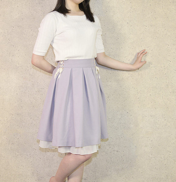
フレアスカートは、春のトレンドのレディースファッションとして注目されています。お店に足を運べば、花柄やドット柄など様々なデザインや色合いの商品を見かけるものです。ここでは、フレアスカートの魅力などについて紹介していきます。
『フレアスカート』とは、身に着けた時のシルエットが朝顔のように広がったスカートのことを指します。「flare」という言葉の通り、生地が大きく広がり、波のような形状の裾が美しく揺れる優雅さがあります…
フレアスカートを上手に着こなす重要なポイントの一つがタックインです。タックインは押し込むという意味の通り、シャツやブラウスといった上着の裾部分をスカートのウエストに入れるファッションになります。フ…
ファッションの印象を左右する要素としては、カラーや素材のほか、デザインも挙げられます。フェミニンな雰囲気に仕上がるデザインのボトムスを選びたいなら、フレアスカートに注目してください。フレアスカート…
ツイードのベストも春のトレンドのレディースファッションとして人気を博しています。オシャレにツイードのベストを着こなすためには、どのような点に注意すればいいのでしょうか。ここでは、ツイードベストの着こなしのポイントをお伝えしていきます。

ツイード素材のアイテムは、春のトレンドのレディースファッションとして注目されています。お店に足を運べばツイードのジャケット、スカートなど色々な商品がありますよね。その中でも特に人気を集めているのが…
ツイードベストは、その素材やデザインから上品な雰囲気を持っています。そのため、着こなしにおいてはシンプルなトップスとの組み合わせがおすすめです。まず、シャツとの組み合わせについて考えてみましょう。…
ツイードベストは、カジュアルからフォーマルまで幅広いシーンで着用できます。着用するシーンによって、素材やデザインを選ぶことが重要です。まず、カジュアルなシーンでは、リラックスした雰囲気を演出するた…